Before you make any decision or take any action based on radiographs (a radiograph is the picture you get from an X-ray examination), you must get a second opinion on what the radiograph really shows. Misread radiographs are very common, and many veterinarians don’t know enough about coffin bone rotation to give you a good understanding of what really has happened.
Picture evaluation costs USD 49 for each picture I evaluate.
A. Email the side-view radiograph to pictures@equinehooves.com, and I will do the evaluation as follows and email it back to you.
I will tell you how to pay through PayPal before I email you the evaluation.
This is what you would get from me:
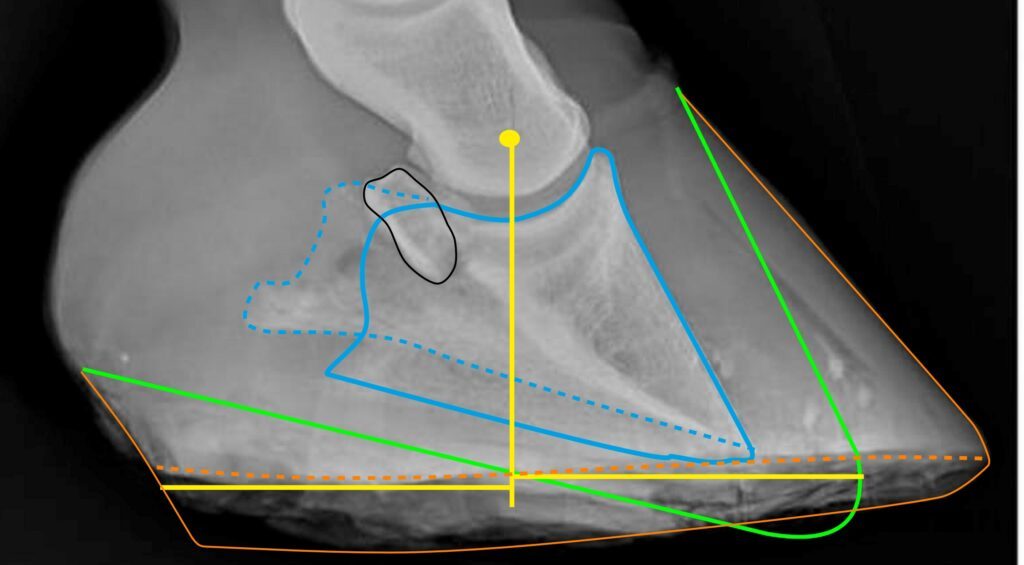
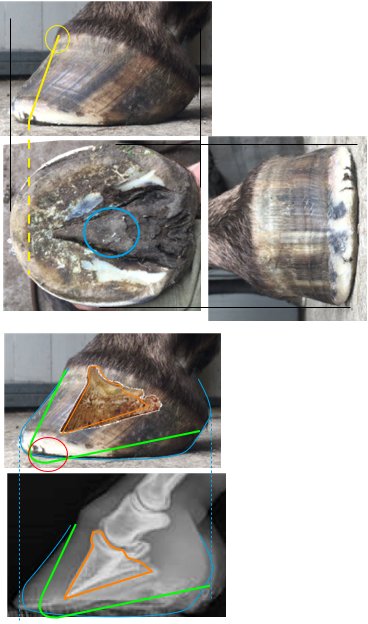
- Orange line = close side of hoof capsule. If it is dark at the bottom of the heel, it means that the hoof wall is too porous to show up on the radiograph.
- Orange dotted line = far side of the hoof capsule.
- Gren line = The hoof shape we want. Now the heels are too high and the toe too long.
- Blue line = close side of the coffin bone
- Blue dotted line = far side of the coffin bone
- Yellow dot = the point from where the horse’s weight loads the coffin bone
- Yellow lines show how the weight is distributed in the hoof. 50% of the weight goes to each side of the vertical line. This means that the heels are overloaded, since 50% of the weight is concentrated on a smaller surface area, which leads to higher pressure per unit area.
The reason the blue lines are different is that the X-ray was taken from too far back and too high.
The bone marked in black is the navicular bone.
This hoof suffers from what they call “Coffin Bone Rotation,” which only means that the hoof wall is no longer parallel with the coffin bone. This is easy to rehabilitate, but NOT the worst problem for this hoof. More complicated to rehabilitate is the fact that the hoof care provider has trimmed away too much under the tip of the coffin bone. This is complicated to rehabilitate, and not all hoof care providers will manage this.
B. Add a photograph of the same hoof. Place the horse on an even floor and place the camera on the floor 2 feet (60 cm) from the middle of the hoof. Make sure the lighting is good.
I will then:
- Adjust the size and position of the two pictures, and draw all the same lines on the photograph as I have done on the radiograph.
- Add relevant comments and email the evaluation to you.
C. Add a photograph of the sole view of the same hoof. Hold the camera 1-2 feet (30-60 cm) perpendicular to the sole. Make sure the lighting is good.
Add one more complimentary photograph of the sole from a slightly different angle. This is to make it possible for me to compare heights.
I will then:
- Adjust and draw lines combining all three pictures. This will give you far more information, and it will be much easier for you to understand.