Equine Physics is not a trimming technique or a hoof care method; It describes the Physics and the Laws of Nature that all techniques and methods must apply to because if physics doesn’t apply to horses, you can be sure they would fly.
Purpose
This segment is written to give you the knowledge you need to really understand what you see in real life and in radiographs. The hoof world is completely roled by opinions and traditions, but if you really want to understand hooves, you must be able to see through traditional misunderstandings and misconceptions about hooves and coffin bones. You need to know where things originate and what are just side effects. As long as the hoof world keeps focusing on compensating instead of removing what is wrong to make room for what is right, hooves will keep being the most expensive part of the horse.
Basics
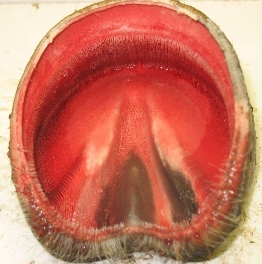
The coffin bone is the centre of the hoof, and everything else must be considered relative to the coffin bone. The coffin bone is where the weight originates and from where the hoof shape is determined. The hoof wall is nothing more than a protective fingernail doubling as a flexible shell for the blood pump. The sole is the primary weight-bearing tissue, doubling as the bottom of the pump enclosure. The frog is the main shock-absorbing landing pad with an essential role in blood pumping, friction on slippery surfaces, and transmitting sliding sensations to the balance centre. The wall, sole, and frog are all tissues made of horn, which means that they contain no blood and no nerves. Blood and nerves are located in the corium to which the horn is connected for stability.
Coffin bones are individual, and the only common denominator is that horses in the same weight category have the same size coffin bones. In physics terms, this means that all coffin bones strive to have the same load per surface unit. Hoof shapes are never breed-dependent, but most often hoof care-dependent. Farriers often believe that a thoroughbred with big hooves will run faster on a soft race-track since a big hoof doesn’t sink into the artificial footing as much. No one recognise that Arabian thoroughbreds, which usually are allowed to have small hooves, are bred for running in loose desert sand?
Hoof care providers always determine the hoof shape. Often lack of symmetry depends on whether they are left or right-handed. If the bar on one side has been left too big because it is hard to reach with the good hand, there is usually a flare on that side. Ground pressure has then pushed that bar to the side, which has made the whole hoof asymmetric. Since this segment is not about bar trimming, I urge you to pick up the next asymmetric hoof you see to compare the bars, and remember what you see until you read the segment on bar-trimming.
An asymmetric hoof with one bar left too long and therefore tilted.
In a laminitic hoof, the pain becomes a problem during the deformation of the hoof capsule. It has nothing to do with a rotating coffin bone, only with a hoof wall being torn loose from the corium. When ground pressure pushes on a loose hoof capsule, it breaks away even further, creating more pain. The outer parts are then pushed upward and outward, stretching, bending, and tearing sensitive tissue inside the hoof.
As long as the force created by the horse’s weight is greater than the ground force, which eventually will stop the hoof from sinking into the dirt, the hoof will keep sinking. When the dirt has been compacted, the ground force (pressing up) increases and eventually becomes as large as the weight-bearing force (pressing down), and the hoof will stop sinking.
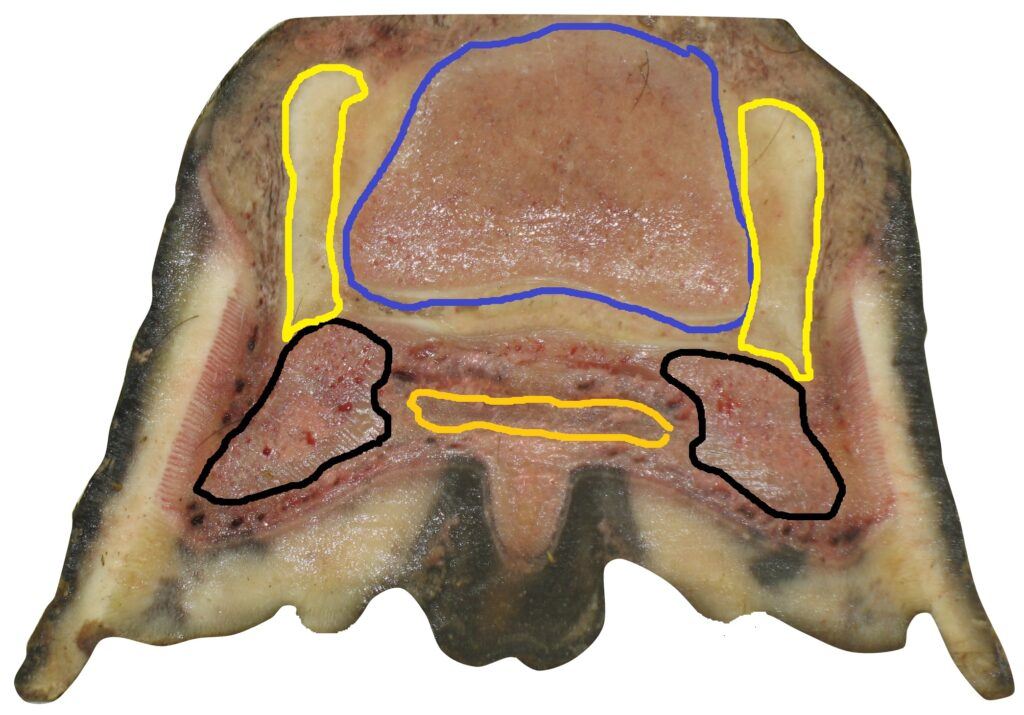
Coffin bone arms = black, Lateral cartilage = yellow, Short pastern bone = blue
When a hoof is being loaded on hard ground, only the hoof wall makes ground contact. The sole directly below the coffin bone will be pressed down, and if it doesn’t make ground contact, all weight will have to pass through the laminar junction to reach the weight-bearing hoof wall.
stretching the sole, and since the hoof wall is the only part of the hoof with ground contact, it will be pressed up by ground pressure.
Ground pressure will stop it from being pulled down by the sole through the laminar junction. When the hoof is loaded on soft ground, what is directly under the coffin bone will be compressed and pushed down into the dirt. What is not pushed down by the coffin bone will be pushed up by ground pressure. The result is a stretched and bent sole.
not be pushed down but rather pulled down by the sole. The coffin bone is standing on the sole (pressing the sole down), but the sole’s connection to the hoof wall (the laminar junction) is stronger than the sole, which will make the sole bend upward. The utmost peripheral part of the sole will be
through the laminar junction, of which none are designed for pulling forces. This creates a pulling, stretching, and bending force in the sole and the laminar junction against the ground pressure (that is evenly distributed under the whole hoof). The peripheral parts of the hoof will be pulled down until the ground force equals the pulling force, when it will stop sinking. This is what causes the severe deformation — and this is where the pain comes from.
A hoof capsule can never be trimmed to be smaller than the coffin bone. When it is allowed to grow larger, problems like deformation, sensitivity, and pain become more severe.
Coffin bones don’t have a standard toe angle, a standard shape, or any standard at all.
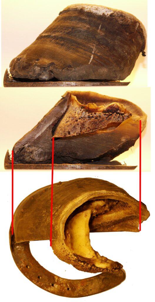
The reason the tip of the lower coffin bone looks strange is that the hoof had high heels for many years. Before we start looking at different coffin bones, I should probably explain why this coffin bone doesn’t look like the picture in the book you have.
You might be used to seeing hooves and coffin bones like this:
Or radiographs like this. If so, let’s hope it wasn’t your horse.
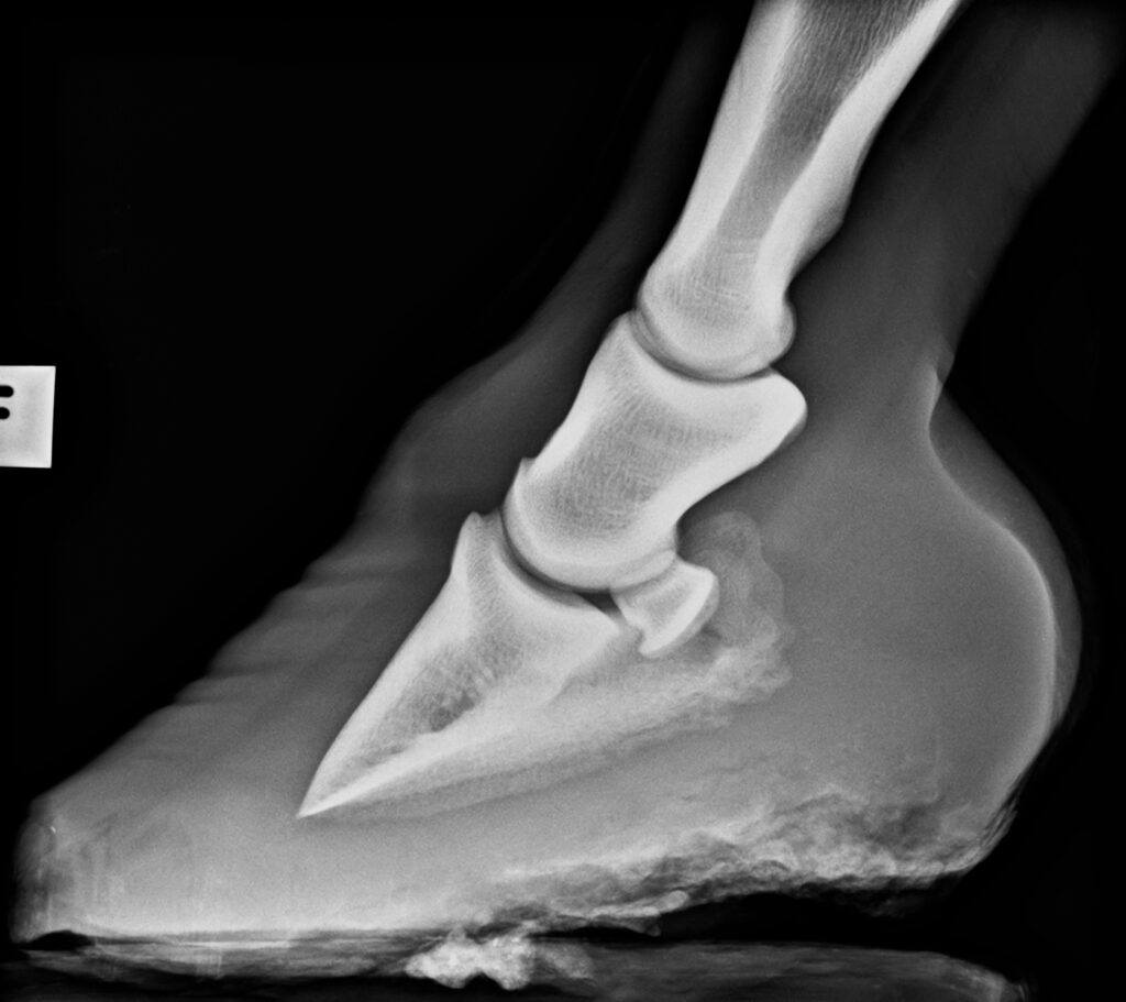
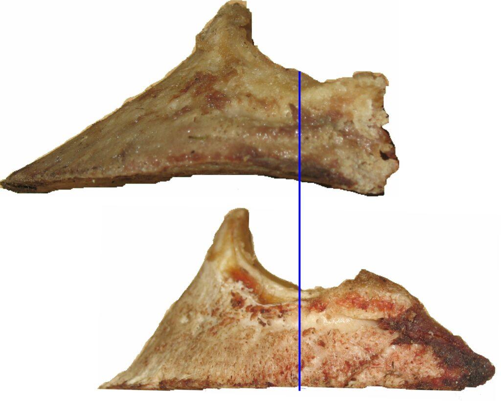
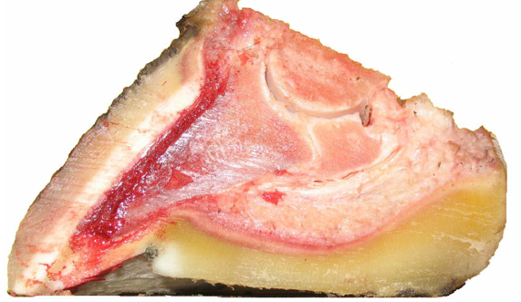
The reason the last two look so different from the first two is that the last two show hooves and coffin bones cut in half.
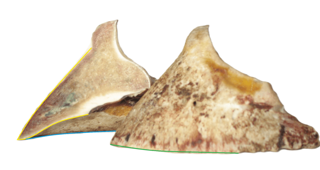
If you cut a coffin bone in the middle lengthwise, place both halves on a table and push the further one to the left, his is what you will see. To the right, you see the close outer surface of the complete coffin bone standing flat on the table. To the left, you see the thickness of the middle of the bone (yellow lines), and the further perimeter (blue line), to the right, you see the close perimeter (green line). The coloured lines are the same in the above andbelow pictures.
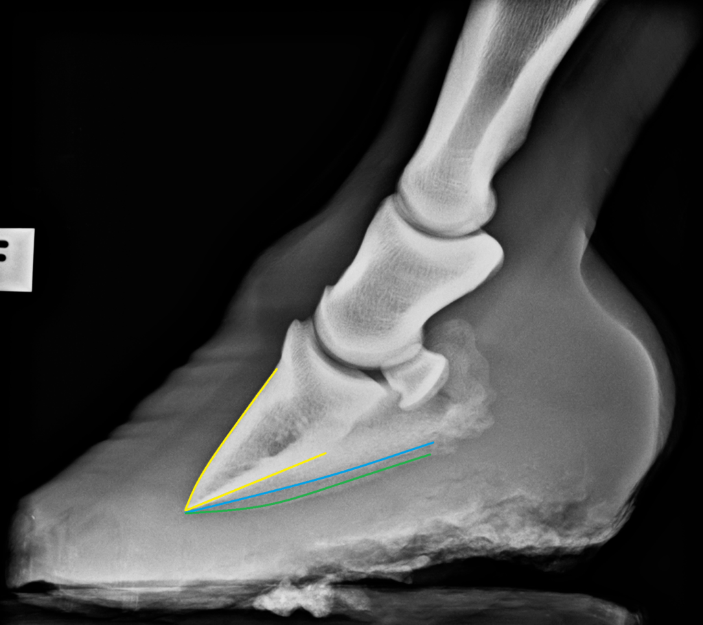
A very common mistake when reading X-rays is to confuse the blue and the green line. The close perimeter is always the lowest line. This is because the X-ray camera was placed higher than the floor and was therefore aiming slightly downwards.
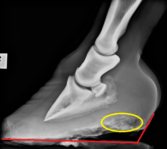
The gray flaming area in the yellow oval is thrush, and the red area just below is hoof wall that was of such poor quality that it did not show up on X-ray (the horse was not standing on his toe)