How can Coffin Bone Rotation be the most fatal condition in the horse world when it isn’t more dangerous than a speed bump, and correct rehabilitation has a 100% success rate?
This page explains why “coffin bone rotation” is not what it appears to be, why it is physically impossible in the way it is commonly described, and why rehabilitation is far more predictable than tradition suggests.
Equine Physics is not presenting a trimming technique or a hoof care method; It describes the Physics and the Laws of Nature that all techniques and methods must apply to because if physics doesn’t apply to horses, you can be sure they would fly.
Table of Contents
- Introduction and Purpose
- The Traditional Explanation of Coffin Bone Rotation
- Anatomy of the Laminitic Hoof
- Why the traditional explanation makes Coffin Bone Rotation fatal
- Why the Traditional Explanation Fails
- What Actually Happens Inside the Laminitic Hoof
- Why “De-rotation” Is a Misleading Goal
- How a Laminitic Hoof Becomes Deformed
- Where the Pain Really Comes From
- Why Shortening The Toe Is Necessary
- Resetting the Coffin Bone Angle
- Realignment of the Toe Wall
- Healing Progress
- Heel Height and Coffin Bone Angle
- After Coffin Bone Rotation Comes Thrush
- Pain from Coffin Bone Rotation or Thrush
- Reading Radiographs Correctly
- A Sound Hoof Hidden Inside the Deformed Hoof
- Why The Tissue Below The Coffin Bone Matters
- Why No Coffin Bone Has Ever Rotated
- The Sole Corium Reveals What Has Happened
- Radiographs show angles, not mechanisms
- Summary of Evidence
- Conclusion
Introduction and Purpose
If you think it is more important to find solutions that make things easy to explain, prevent, and rehabilitate than to preserve an old tradition that leads to incorrect explanations that break the laws of physics and cause suffering and pain, then I am convinced you will feel at home here.
“Coffinbone rotation” is the deadliest condition in the horse world. It is, however, only a theory that has never been proven, and it is based on an assumption that defies physics, so it can’t be correct. Of course, I’m not saying that something isn’t seriously wrong in a hoof with coffin bone rotation, or that those poor horses don’t suffer excruciating pain. I’m just saying that with the right explanation of what has happened, it becomes easy to both understand and rehabilitate. The objective of the Good Protocol is to make the knowledge foundation needed for 100% successful rehabilitation of coffin bone rotation available to everyone.
You only need to know two things;
1. Where does the pain come from?
2. What causes the pain?
You don’t need years of specialised education or extensive experience.
The objective for this segment is to explain how the theory of the condition coffin bone rotation is defined, how it is explained, and why it is physically impossible and would break the laws of nature. You will also gain the knowledge to understand what really has happened, where the pain comes from, and how to avoid it. If you have traditional knowledge about hooves, it will be easier for you if you choose to put everything you have heard earlier about hooves aside and enter with an open mind. I intend to give you evidence for everything I say that contradicts the traditional opinion.
The Traditional Explanation of Coffin Bone Rotation
The definition of the condition “Coffin Bone Rotation” only states that if the toe wall and the coffin bone are not parallel then the hoof has the condition. The condition says nothing about what has happened just what is. To this, The Good Protocol agrees completely. If the hoof wall and the cofin bone are not parallel that hoof suffers an issue that might be called a condition. It is the choice of name and the traditional explanation for what has happened that makes the condition lethat, not the condition.
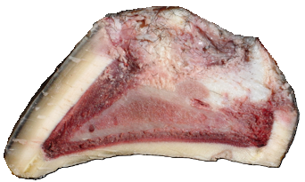
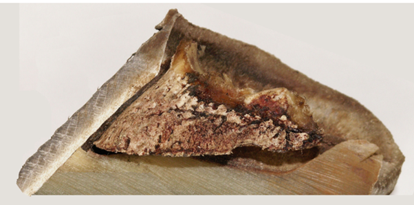
All horse books with a chapter on coffin bone rotation show the same basic picture. There is nothing wrong with that picture, except that it doesn’t show you what the inside of a hoof looks like:
- You don’t see the shape or outer form of anything, just what they look like cut in half.
- That picture doesn’t show anything interesting about the coffin bone.
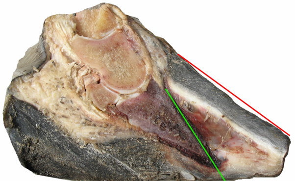
The picture doesn’t lie. It shows clearly that the toe wall and the coffin bone are not parallel, and yes, that is what most people look for, since that is what the horse world’s most fatal condition, “Coffin Bone Rotation,” is based on.
Since a picture like this generates questions, they had to come up with explanations and here is where the ltotal ack of science based knowledge shines through.
The traditional explanation to what caused the condition is that the coffin bone was suspended by the laminae parallel to the hoof wall we can see in the picture and, when the hoof suffered laminitis, the Deep Digital Flexor Tendon (DDFT) pulled hard enough to rotate the coffin bone down to where it is in the picture.
The traditional treatment reasoning is that since he coffin bone has been rotated down it needs to be rotated back up again for the hoof to become healthy
From here on let us add science and physics to this and let us see where that leads us.
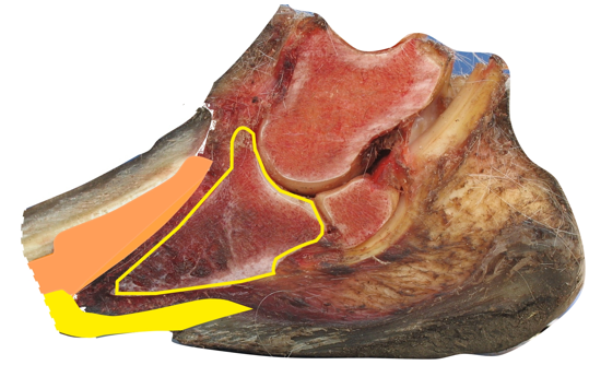
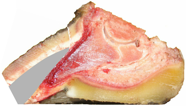
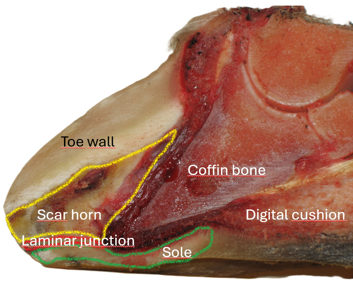
Anatomy Of The Laminitic Hoof.
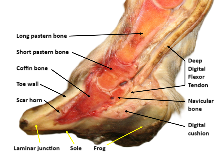
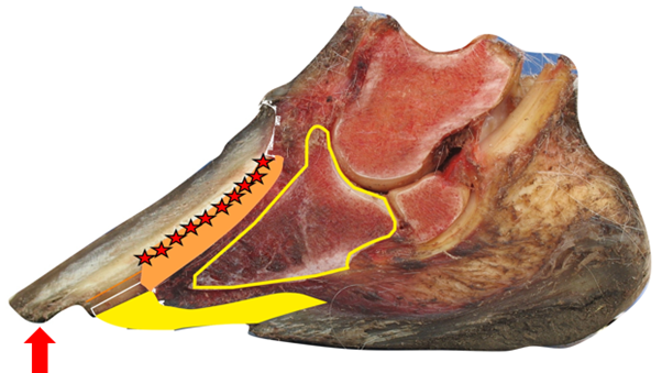
I assume you are familiar with most of what this picture shows but let me explain two things:
- The laminar junction is the very strong connection between the sole and the hoof wall. This tissue is called many things, like “white line”, but it is not white and if we go back in history, the while line was distinctly defined as the thin, crisp white inner layer of the hoof wall.
- Scar horn is wound serum that has hardened to a dense material without blood or nerves. It is very well connected to the primary lamella and will follow the hoof wall wherever it goes.
The scar horn filling the void between the hoof wall and the coffin bone makes it impossible to push the coffin bone back up to its alleged original position by the hoof wall, since that space is no longer vacant. The void is said to have been created when the coffin bone was rotated down from its alleged natural position hanging parallel to the hoof wall suspended in mid air by the laminae, to where we see it now.
The traditional answer to how the coffin bone was rotated down from the hoof wall is that laminitis impaired the laminae connection, suspending the coffin bone, which allowed the pull in the Deep Digital Flexor Tendon (DDFT) to rotate down the coffin bone from where it hung, tearing the laminae apart. When the void between the hoof wall and the coffin bone was created, it was immediately filled with wound serum, which hardened to scar horn. That is how the traditional explanation of Coffin Bone Rotation makes the condition lethal, since this can’t be treated, cured, or rehabilitated.
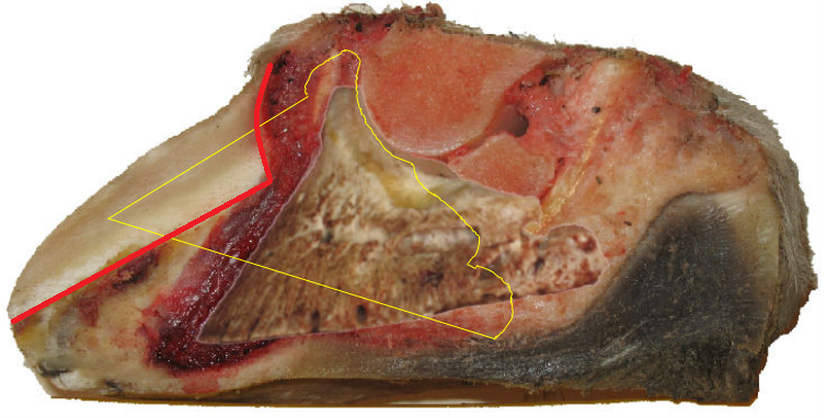
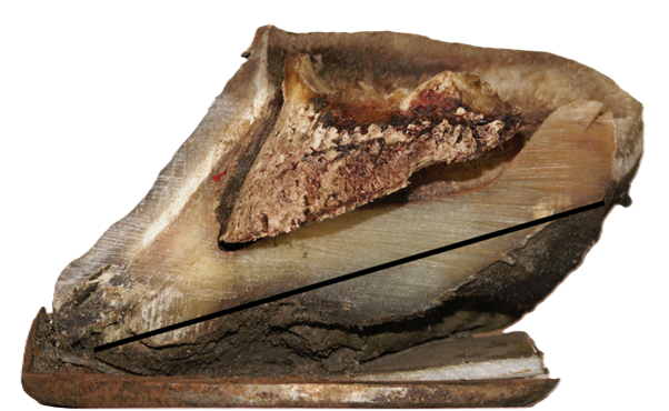
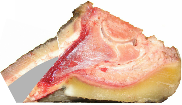
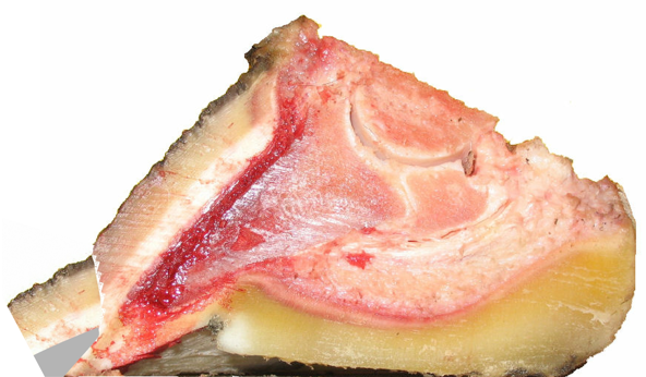
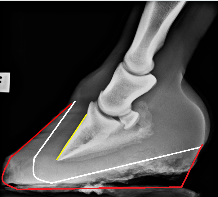
When we place the sawn-off half of the coffin bone where it belongs, we can see that this hoof has the condition “coffin bone rotation” (the hoof wall and the coffin bone are not parallel) even though the coffin bone is almost completely parallel to the ground. If the coffin bone is parallel to the ground, it can not be rotated. Most people don’t have access to the other half of the coffin bone, so they can’t see its angle to the ground.
If the coffin bone toe is a degree or two higher or lower, instead of being perfectly ground parallel, it is of no importance since the ground is seldom perfectly horizontal anyway. I will write a separate segment on the misconception called NPA later.
If a coffin bone were to rotate, it would have to rotate around the ball joint. When doing that on this picture, we get a result that no one can believe is natural. The coffin bone would have had a very unlikely angle to the ground, and it would have been placed where the hoof wall is today. Too bad they didn’t have access to the other half, because if they had, the condition coffin bone rotation would probably never have existed. This is because a coffin bone that was parallel to a deformed hoof wall before being rotated down most often would not fit in the hoof capsule.
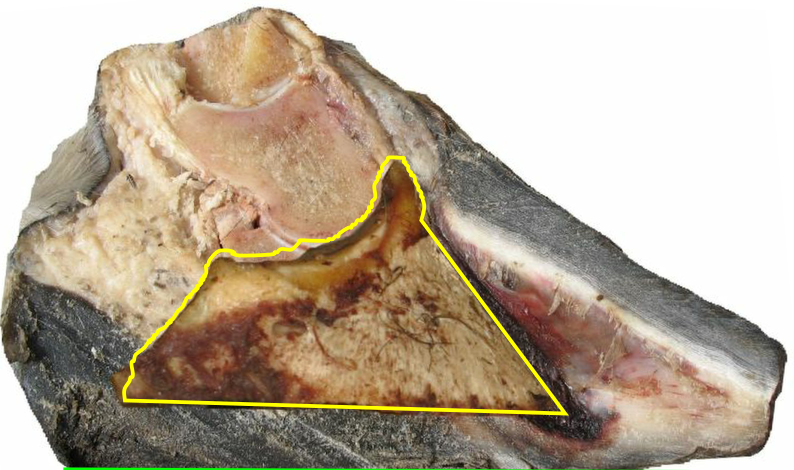
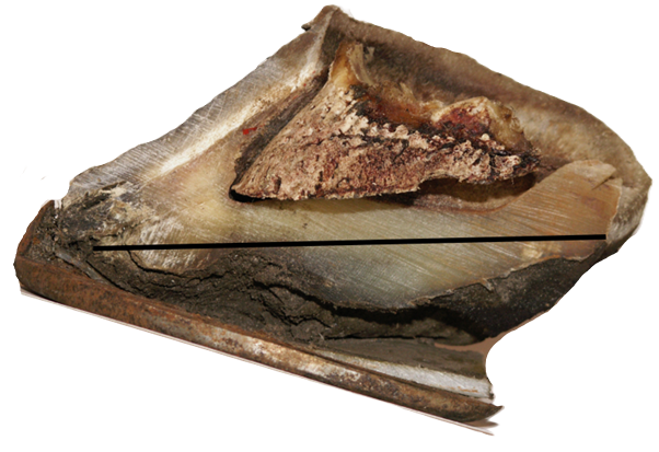
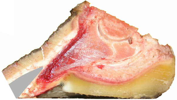
It is not this extreme on all laminitic hooves, but it is too common to neglect. Let me show you another one. If the front of the coffin bone had the same angle as the toe wall before the alleged rotation, it would have been placed as the yellow marking shows.
To prove that I’m not cherry-picking the pictures, I will show you a radiograph where this is not as obvious later. All the evidence does not need to be obvious on all specimens when one is enough by itself.
Now the choice is yours: trust the tradition and accept the consequences, or read on, and the Good Protocol will prove to you that the condition Coffin Bone Rotation doesn’t need to be more than a speed bump, and the only reason for rehabilitation to fail is ignorance.
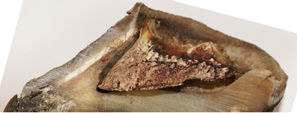
This picture, showing a hoof with the condition Coffin Bone Rotation, is only here to show you the big picture. There will be many other pictures showing the details during the rest of this segment.
Why the traditional explanation makes Coffin Bone Rotation Fatal
Traditional rehabilitation is based on that the coffin bone has rotated down from its original position parallel to the hoof wall to where we can see it on the radiograph. To rehabilitate the condition, they want us to push the coffin bone back up where they say it belongs. This is, however, impossible since that space becomes filled with scar horn as soon as it is vacated. Scar horn is a dense material without any blood or nerves in it. The only way to get rid of scar horn is to wait until the hoof wall moves it down to the ground. As long as the toe wall stays too long and there is ground pressure affecting the laminitic hoof wall, the hoof wall will never retake its natural angle, and new scar horn will continuously be produced. As long as you believe that the coffin bone must be de-rotated, the condition stays permanent and therefore fatal.
Why the Traditional Explanation Fails
The traditional explanation fails to account for several well-known observations:
The DDFT cannot rotate the coffin bone because the attachment point of the Deep Digital Flexor Tendon is located close to the middle of the coffin bone, not near its tip. This means that, for the tendon to rotate the bone downward, the force required would have to be unrealistically large.

In addition, the DDFT is not under constant tension. It is activated during the push-off phase of the stride — a phase that laminitic horses actively avoid because it is painful. A mechanism that requires active push-off cannot explain a condition that occurs in horses that are barely willing to load the limb at all.
This alone makes the traditional rotation mechanism mechanically implausible.
- For any structure to carry hanging weight efficiently, it must be aligned with the direction of the force it is intended to transmit. The laminae are not. The laminae are angled perpendicular to the hoof wall, which tells us that they are meant to hold the hoof capsule, sometimes to the left, sometimes to the right, and sometimes holding the capsule down. Not a single lamina in this cross-section is therefore meant to carry vertical load.
This physical evidence alone makes the traditional rotation mechanism mechanically implausible.
Since the coffin bone can’t hang from the laminae, the horse’s weight must follow a different load path through the hoof.
This does not yet tell us what carries the weight — only that the traditional suspension model cannot be correct. There will be a complete segment on the weight’s way through the hoof. That will be an interesting one, don’t miss it.
- Removing the condition doesn’t imply derotation of the coffin bone. By rasping the toe wall to the red line, the condition will disapear but nothing has changed inside the hoof. I have seen this done both in books and in practice, sometimes accompanied by a radiograph proudly showing that the “rotation is gone.” This is not rehabilitation.

Parallel does not mean healthy
In a sound hoof, the hoof wall is parallel to the coffin bone, and the coffin bone is parallel to the ground.
Just because the hoof wall and the coffin bone are parallel doesn’t mean the hoof is sound.

A hoof where the toe wall and coffin bone appear parallel can still be severely compromised. The crucial question is not whether these structures are parallel, but which part of the hoof has changed position to create the observed relationship.
This distinction is not academic. In the rehabilitation of laminitic hooves, it is often a matter of survival.
A ground-parallel coffin bone can be diagnosed with “rotation”, and a hoof with any shape can be diagnosed as not having coffin bone rotation as long as the hoof has the sought-after parallellity.
Since the condition Coffin Bone Rotation is only defined by the coffin bone’s and the toe wall’s parallelity, the coffin bone can have any angle to the ground and still be diagnosed with Coffin Bone Rotation

On a hoof with a ground parallel coffinbone it is always possible to trim away the condition without affecting the coffin bone in any way. It might be considered aggressive since scar horn is not as protective as real horn, but it shows the principle of effective rehabilitation.
There are signs that you can learn for how much toes can be trimmed while staying on the safe side, and they have nothing to do with the so-called “white line”. On a hoof with high heels, which always creates a tilted coffin bone, the heels and toes must be addressed in tandem for effective and safe rehabilitation. Trimming laminitic hooves is nothing out of the ordinary and doesn’t require any special education, just thoroughness. The same cannot be said about hooves that are close to the coffin bone, penetrating the sole, like the hoof below. Such hooves are very challenging to rehabilitate.
Trimming a high-heeled hoof to get rid of the coffin bone rotation would give the hoof an unnaturally steep toe, but trimming the heel and the toe in tandem would remove the condition while keeping a natural toe angle. I will address trimming hooves like this in a segment called rehabbing traditional trims.
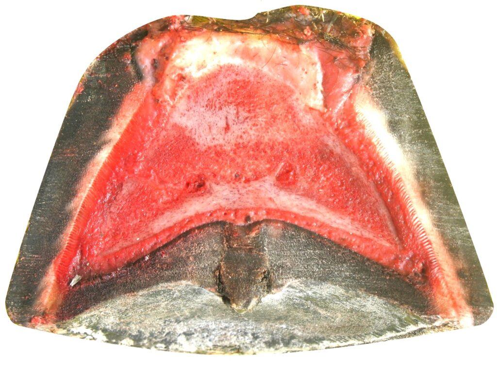
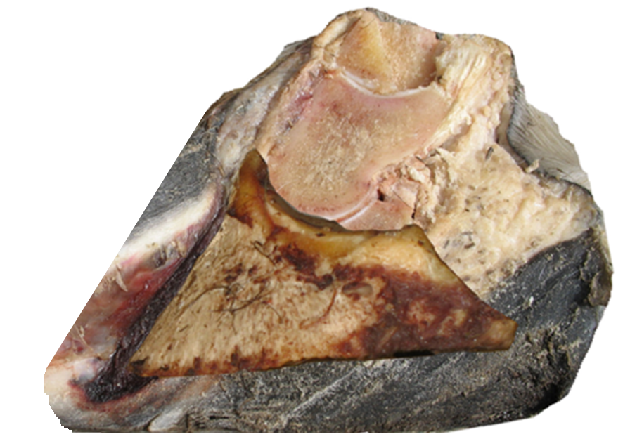
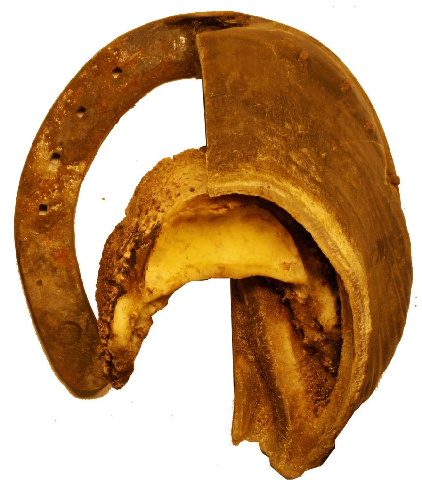
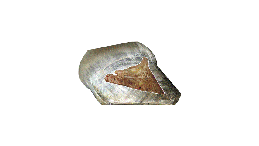
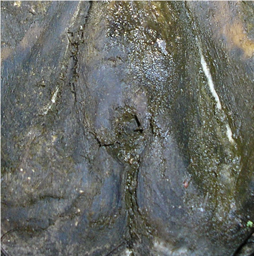
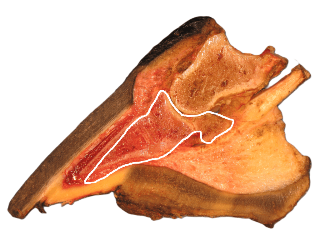
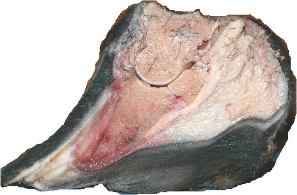
Since the hanging and rotating coffin bone is so central in traditional hoof care, I’d like to show you another picture of what it looks like around the coffin bone. When cutting a hoof lengthwise, but shifted slightly to the side, so it passes through one of the lateral wings of the coffin bone and therefore shows a longer coffin bone than when the coffin bone has been cut in the middle. This makes the coffin bone look different from the earlier picture, but both images are correct. I’d like to point out that the hoof in the picture doesn’t suffer from either laminitis or coffin bone rotation. This is what a sound hoof looks like inside.
The sole corium has an even thickness of approximately 1/8”= 3–4 mm, and under the corium comes the sole, and directly under the sole, there should be the compressed ground we can see in hoofprints. There is no space anywhere for the tip of the coffin bone to rotate downward into. If the tip of the coffin bone were still to rotate down, it would crush the corium below it. There is no way of checking the condition of the sole corium on a living horse, but I have never seen a crushed corium in any dissected hoof.
These inconsistencies do not yet tell us what has happened — but they make it clear that the traditional explanation cannot be correct.
To understand what is actually going on inside the hoof, we must move beyond assumptions and examine physical constraints and anatomical reality.
What Actually Happens Inside the Laminitic Hoof
This video shows how coffin bone rotation is created.
https://www.youtube.com/embed/ptTvFUvJE90?feature=oembed
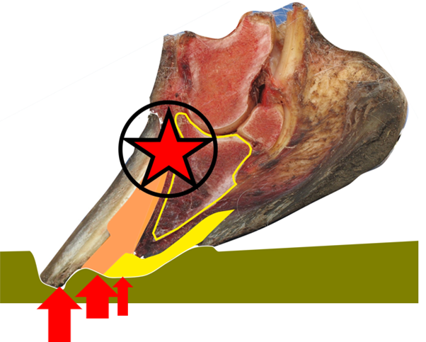
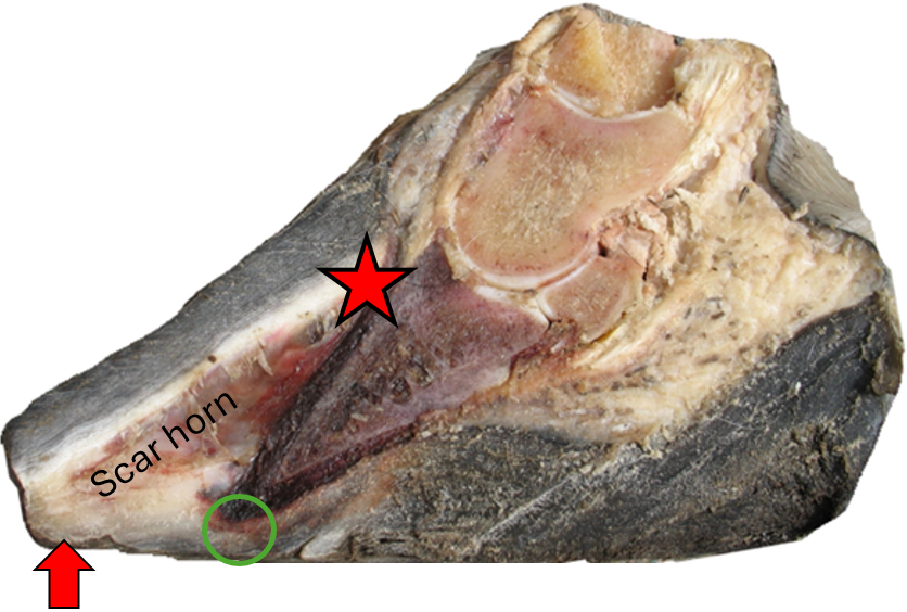
When the hoof is affected by laminitis and the number of functioning anchoring cells, which are supposed to hold the hoof capsule to the corium, is reduced, the hoof capsule becomes loose and easily deformed by ground pressure. If the toe wall is then pushed upward by ground pressure (the red arrow), lamellae will be torn loose from the corium — under horrific pain. This can even happen without the laminar junction (the connection between the sole (yellow) and the hoof wall) breaking.
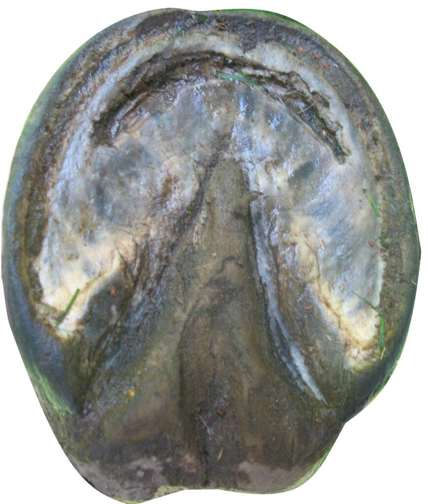
On a hoof with a sound laminar junction, but a too-long toe before foundering and getting Laminitis, the ground pressure might push the loose toe wall away from the coffin bone until the sole broke. On high-heeled hooves, such issues can lead to the condition of sole penetration and death. The picture below shows a stretched sole, which has nothing to do with the angle of the coffin bone or the bone penetrating the sole, but only a loose hoof wall that has been pressed up, pulling and bending the sole around the outer edge of the coffin bone enough for the sole to be stretched thin and break.
The new void created when the lamina was broken loose from the corium fills with wound serum, which later hardens into scar horn.
Every time the toe becomes exposed to ground pressure, more of the still-functioning attachment between the toe wall and the coffin bone tears apart under unbearable pain (red star).
It is the long toe that creates the lever forces that make ground pressure destroy the corium and cause the pain.

With a defective connection between the hoof wall and the coffin bone, breakover becomes intolerable, and this is why laminitic horses take short, careful steps — they must lift the hoof before the toe is subjected to ground pressure.
There are signs you can learn to read to be sure exactly how much you can shorten the toe, and I will come to them in a separate segment. If you have the knowledge, you can shorten the toe all it needs in one trim, but if you don’t, I recommend that you take it little by little to see when he can walk again and then keep the hoof like that with daily trims. Doing nothing is not an option. The best way to rasp a hoof like this is vertically downwards, not to irritate the very sensitive corium. There is a way to do this trim even if you can’t lift the hoof, but I don’t want to talk about trimming details yet. I’ll come back to that later. Trimming laminitic hooves is essential for quickly reducing unnecessary pain.
Since the whole problem that is called “coffin bone rotation” is caused by ground pressure against a laminitic toe, shortening the toe is when the magic happens. The effect is instant. As soon as ground pressure is no longer affecting the toe, the lameness goes away, and the horse wants to gallop. Some horses might need up to 20 minutes before they gallop, and if so, then it is the “pain memory” that makes the horse not dare to try, but when they have walked for a while, they realise that it doesn’t hurt anymore, and they can’t stop themselves from galloping. Of the over 1000 laminitic horses I have trimmed, not a single one has not been galloping within half an hour. Quite often, they have done 10 laps around their paddock, bucking, and their owner has cried with happiness. If the horse doesn’t gallop after the trim, the toe is still too long, or maybe the heel is too high. A high heel tips the hoof forward, which makes it easier for ground pressure to reach even a short toe, so the correct trim must be a combination of shortening the toe and lowering the heel.
Why “De-rotation” and “Re-rotation” are Misleading Goals.
There are no internal forces or swelling connected to Laminitis that can rotate the coffin bone or push out the hoof wall. The weight of the horse presses the short pastern bone down through the hoof joint to the coffin bone (blue arrow below). If the coffin bone had been suspended by the laminae of the hoof wall, which is connected to the corium (reddish marking) on the coffin bone, physics says that the horse’s weight would be pressing down the back part of the bone harder than the front part, which they say is being rotated down. None of this is, however, an issue since a hanging coffin bone would break the laws of nature. You can read more about this in the segment on Laminitic hooves. The coffin bone is always standing on the sole, never hanging in the air from the hoof wall, the DDFT is relaxed when the horse is standing still (look it up in any book on horse anatomy), and there is no force pressing down on the tip of the coffin bone. This means that no forces are pushing or pulling down the tip of the coffin bone on a standing horse. This should be fantastic news for the horse world since it means that coffin bones don’t rotate, and shortening a long toe is much easier than pressing up a coffin bone that has never rotated down.

The evidence that this is correct lies in the trimming, so try it. I have done it thousands of times with 100% successrate. Any hoof care provider can shorten a toe. A little later, I will tell you how much you can shorten the toe, and even tell you how to shorten a toe without lifting the hoof. Waiting for the veterinarian to come and give the horse a painkiller instead of shortening the toes on a laminitic hoof must be considered animal cruelty. This is a tradition that must be changed.
How a Laminitic Hoof Becomes Deformed
The horse’s weight travels in the skeleton all the way down to the coffin bone, and from there through the sole corium, and the sole to the ground. Without this, the sole corium would not get any blood circulation. The force of the ground pressure must equal the gravitational force created by the horse’s weight, or the horse would keep sinking into the ground or fly up in the air. When the hoof has stopped sinking into soft ground, the ground pressure is distributed fairly evenly over the complete footprint. This is, however, probably a little scarier than you think because it is only the part directly under the coffin bone that is being pushed down. The rest of the hoof print, which is larger than the coffin bone, will instead be pulled down by the sole and the laminar junction. The coffin bone pushes on the sole, and the sole pulls the hoof wall down in the dirt, and all this force must pass through the laminar junction. This means that the outer edge of the originally arc-shaped (concave) sole will be stretched upwards, first pulling the sole flat and later even U-shaped (convex). Commonly, the rounded shape of the sole becomes misinterpeted for the sole being swollen, but the sole horn can’t be swollen, no horn can. When something becomes swollen, it means that is absorbes more water, but horn can’t absorb enough water to make it visibly swollen.
To make this easier to understand, think of a gentleman placing his jacket on a mudhole for the lady to step on without being muddy. When she steps in the middle of it, her foot will push down the middle of the jacket, and the jacket under her foot will gradually pull down the rest of the jacket into the mud. Since ground pressure is pushing up on everything that is still horizontal, the parts of the jacket that are furthest away will stay on top of the mud longest, but gradually be pulled toward the middle.
This means that the part of the hoof’s sole that is under the coffin bone will be pushed down, and the peripheral parts of the sole and the hoof wall will be pushed up by ground pressure, but gradually pulled down by the sole. Since the toe wall is angled out, a vertical force pointing straight up will push the lowest part of that hoof wall out and away from the hoof and the coffin bone. While this happens sole will be stretched and bent upwards in a completely unnatural way around the outer edges of the coffin bone. The larger the diameter of the sole is, compared to the diameter of the coronary band, the greater the leverage and the deforming forces will be. This is very bad news for those who believe that hooves are supposed to be big, even the bigger the better. That is painfully wrong. A small hoove give much less leverage for the ground pressure to deform the hoof compared to a large hoof. If a hoof has been allowed to grow bigger than the coffin bone, that hoof will have “coffin bone rotation” or “distal descent” (lowering of the coffin bone), or both, since the wall has been pushed out (coffin bone rotation) and/or up (distal descent).
If the coffin bone is lowered in the hoof capsule (distal descent) or the hoof capsule is being elevated compared to the coffin bone (no condition) might sound semantic, but the seriousness of the difference becomes obvious when you try to rehabilitate it. If you try to push the coffin bone back up in the hoof capsule, you will just lift the whole skeleton; the enlarged measurement between the upper tip of the coffin bone and the coronary band will be consistent, and nothing will change in the hoof. If you instead unload the hoof wall, the hoof capsule will retake its natural position by itself.
The ground pressure affects the hoof differently depending on where the force is applied. Ground pressure on the frog should never be a problem, but often is, but then it is because of thrush (see separate segment). Ground pressure on the sole directly under the coffin bone is not a problem, but the further away from the corium (yellow marking) it’s applied, the greater the leverage becomes, and with that comes a greater damaging force. Laminitis makes this problem much worse since it incapacitates the laminae that are supposed to keep the hoof wall well connected to the coffin bone.

Where the Pain Really Comes From
The excruciating laminitic pain comes from ground pressure pressing the peripheral parts of the sole and the hoof wall up from their natural positions, leaving a severely deformed hoof capsule with torn laminae. There are no nerves in either the hoof wall or in the scar horn, so even though the pain comes from the lamina being torn loose from the corium, it is the corium side that hurts. When the lamellar attachment between the hoof wall and the coffin bone is weakened, it can no longer withstand the bending and breaking forces created as ground pressure pushes the hoof wall away from the corium, which is firmly attached to the coffin bone.
The remaining healthy attachment between the hoof wall and the coffin bone is then torn apart under excruciating pain as the toe wall is leveraged away by ground force pressing on long toes.
As the toe wall is pushed upward and torn away from the coffin bone, the resulting cavity fills with serum, which later hardens into scar horn. The scar horn is attached to the primary lamella on the inside of the hoof wall.
If any pain comes from the sole corium beneath the tip of the coffin bone (green marking) depends on two things: the coffin bone’s angle to the ground, which we can’t judge without seeing the shape and angle of the whole coffin bone, and whether the sole has been stretched thin and bent around the tip of the coffin bone. The only thing that can make the tip of the coffin bone point into the sole is incorrect trimming. There are no natural forces that can make the tip of the coffin bone penetrate the sole in a naturally shaped hoof.
Why Shortening the Toe is Necessary
Usually, shortening the toe wall is sufficient for a laminitic horse — moments earlier unable to take a single step due to pain — to gallop away willingly and happily. If the heels are high, lowering them helps reduce load on the toe wall further, especially during breakover.
The laminitic pain disappears immediately when the toe wall is shortened enough to eliminate ground pressure on it. You can reduce pain by trimming to the “white line*,” but that is hardly ever enough to lead to recovery. You must change enough to trigger the transformation that will lead to rehabilitation. Taking too little will not make the hoof aim for full transformation, and you can keep doing little forever without getting anywhere. Take enough, and the hoof will focus all energy on transforming as fast as possible. This means that if you lower the heels enough, i.e., take all that the hoof shows you is okay, He might ask you to take 4cm=1.5” more in two weeks, or less. If the hoof shows you signs for trimming 4 cm = 1.5”, that is what you do. You don’t get any points for taking less. You just start a process that will inspire the horse to start a transformation of the hoof, and then you will have to remove what the hoof wants to get rid of.
The Proof is that it works.
*The original definition of “white line” is the unpigmented (crisp white), most inner thin line of the hoof wall. Today, it seems like everyone has their own definition of “the white line,” and that is seldom even white.
Shortening the toe vertically in accordance with the Good Protocol is never a bad thing. Even though you might know that the hoof is laminitic and a shortened toe would solve the lameness problem, you can’t be sure that it isn’t suffering from something else at the same time, and that might be more difficult to rehabilitate.
Having the toe shortened from underneath to create a steeper toe is among the worst things that can be done to an equine hoof.
Resetting the Coffin Bone Angle
The only thing that can affect the angle between the coffin bone and the ground is the relation between the height of the heel and the height of the toe. These two must be considered in tandem to balance the coffin bone. If the tip of the coffin bone is pointing downwards, the heel is too high, and if the tip of the coffin bone is too close to the ground, the front part of the hoof has been trimmed from underneath.
There is very seldom a need for a radiograph to determine the perfect heel height. There are external signs you can learn to read for how much you can lower the heels, so you don’t have to guess, and if the result would be a degree or two more or less is of no importance since the ground is hardly ever perfectly horizontal.
When the heels are readjusted to natural height, the coffin bone follows exactly. It has no other choice.
The reason for shortening the toe of a laminitic hoof is to make it impossible for ground pressure to come in contact with the toe wall, which would create pain from tearing the corium. In extreme cases, where the toe has been very long, you might have to trim far into the sole since ground pressure to the old scar horn also can push the hoof wall away. I know that trimming into the sole is considered taboo, but with the right knowledge, it can be done safely. I’ve done it hundreds of times without drawing a single drop of blood. I have even guided desperate horse owners remotely with 100% success rate, so those who believe trimming laminitic horses is something only farriers with extra special education can do are not well informed.
Keep the new height of the heels as low as possible and make sure there is no ground pressure reaching the toes until the new hoof wall has reached the ground (in a year or so). As long as you make sure the toe wall is not being pressed out, the new hoof capsule will grow down to its natural shape and position “by itself”.
Realignment of the Toe Wall
Laminitic hooves want to grow completely healthy again, so it’s just a matter of not stopping them. All scar horn is attached to the laminae of the old hoof wall and will grow out — if given the chance.
The new hoof wall produced at the coronary band follows the coffin bone downward, as long as it is not forced outward by ground pressure against the old toe or the scar horn.
So keep the toe short.
Be sure to always rasp the toe vertically and only push the rasp downwards to not cause pain and destroy the hoof by breaking the hoof wall outwards.
When the healing angle between the old and new hoof wall has grown down to roughly half the hoof height, it is common for horses to suddenly become extremely lame. This is usually because the lower, old part of the hoof wall is quite loose and moves easily, while the upper, new hoof wall is very rigid. A pressure point can then occur between these two parts, pressing painfully into the sensitive corium. This problem typically disappears instantly when the toe is shortened further. You must learn to ignore all traditional trimming rules, like not trimming past the white line, or you will not stand a chance.
A hoof that has been drastically shortened will often look very different after the trim, but following the protocol, there is no risk of drawing blood. There is no blood in scar horn. There are two different signs indicating how much a toe is too long, and if they say the same thing, they override all traditional limitations, and you can safely trim far into the sole.

When only about 2 cm (1 inch) of old wall remains, it can be very tempting to rasp off the last part to finally make the hoof look neat again. Doing that right does not cause a problem, but be careful not to remove too much. Remember that the primary purpose of the hoof wall is to protect the delicate internal structures.
Hoof wall grows with approximately 1cm/month = 1 inch per 2.5 months, and all new growth is to be considered a replacement of old tissue, not a repair. This means that the new tissue will be just as strong as the original tissue was before the laminitis.
The Healing Progress

Riding can start as soon as the horse (or pony) doesn’t show any signs of lameness. This usually means long before the hoof has regained its pretty shape. There is absolutely no need for a long resting period if the rehabilitation has been done according to the Good Protocol.
If the trimming has been done correctly, the “healing angle” at the top of the hoof wall should determine the angle of the entire new hoof wall.
Heel Height and Coffin Bone Angle
The heel height is the only thing that affects the angle between the coffin bone and the ground, but heel height doesn’t affect the condition Coffin Bone Rotation at all. The coffin bone can only penetrate the sole if the heels are too high and the sole has been thinned from underneath, which is a standard procedure in traditional hoof care.
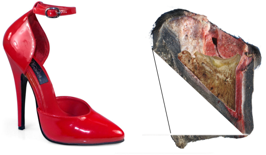
If the coffin bone is not ground-parallel, the horse is essentially wearing high heels, which might look elegant or sexy but is hardly practical in the arena or in the forest. A high heel leads to a tilted coffin bone, which creates an imbalanced hoof. Even worse is that high heels unload the frog, which leads to contracted heels, reduced capacity to read the ground, diminished hoof mechanism and reduced blood circulation in the hoof.
After Coffin Bone Rotation comes thrush
Thrush is a very common and difficult-to-cure issue. First comes the fungus from the inside that kills the cells, and then comes the traditional bacteria that break down dead cells in nature. This impairs the frog, making it less protective, which leads to the heels growing faster to lift the frog away from stones. It is very common for deformed laminitic hooves in the early stages of rehabilitation to develop serious thrush. Therefore, I recommend treating all such hooves preventively. More on thrush will come in a separate segment.
Pain from Coffin Bone Rotation or Thrush.
When a horse stands with the lower part of all four legs pointing forward, he is unloading the toe regions. As soon as his toes have been shortened enough, he will stand straight again.

When the pain comes from the rear parts of his hooves, the frogs, he will stand “under himself” to press down the toes to unload his thrush-infected frogs. No laminitic horse would ever stand like this.

Reading Radiographs Correctly
How can ground-parallel coffin bones get the diagnosis “coffin bone rotation”?
- Because many don’t know what to look for. (They are not looking for the angle between the coffin bone and the ground.)
- Because many people misread radiographs. (They don’t understand what they see.)
- Because many radiographs are not horizontally aligned. (The X-ray device was placed too high.)
- Because many radiographs are not taken perpendicular to the hoof. (The X-ray device was often placed too far back.)
- Because the X-ray device was placed too close to the hoof. This creates a skewed perspective.
A Sound Hoof Shape Hidden In a Deformed Laminitic Hoof.
Hooves suffering coffin bone rotation can look like this. Long toes and high heels. If this is tradition or fashion doesn’t matter. It is very common and very wrong.
With some trimming and some time, the hoof would soon become nice-looking and healthy again. My white line shows the wanted hoof shape, and as you can see, it is absolutely doable. The horse wasn’t standing on his toes when being X-rayed, so the black part under the heels is hoof wall that was of too poor quality to show up on a radiograph (just like you can’t see the digital flexor tendons in this picture (tendons are just not dense enough).
If you understand that the problem is the hoof capsule — not the coffin bone — then “coffin bone rotation” is easy to rehabilitate. There is truly no reason to fail.
Reading radiographs is very different from looking at photographs since the X-ray adds all the tissue it passes through and presents it as one layer, and that is why you can’t determine sole thickness from a radiograph. Even if you know where the sole is, what you see can still be the sum of: hoof wall, corium, laminar junction, sole diagonally, air, sole diagonally, laminar junction, corium, hoof wall, depending on where you look and exactly in what angle the radiograph is taken.
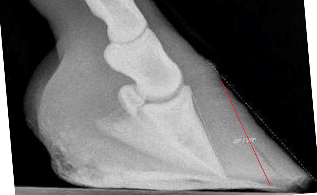
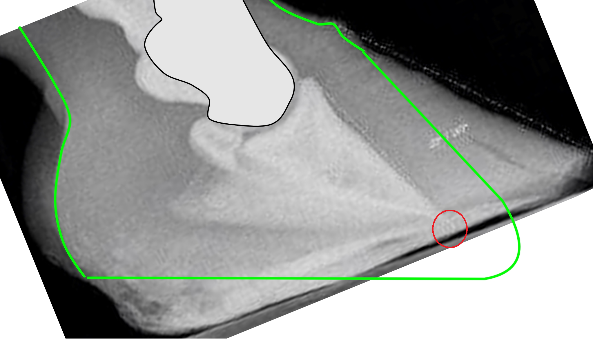
I knew the horse whose hoof appears in the following radiograph, and I know that the hoof got diagnosed with “coffin bone rotation” based on this image (which is why I have it).
If this hoof was anything but sound, it had nothing to do with anything we can see on this radiograph. The toe might be a little too long (like on most hooves), and the hoof wall is not perfectly parallel with the coffin bone (like on most hooves with long toes), but the coffin bone is almost perfectly ground parallel (which is rare). If the coffin bone has an angle of a degree or two, up or down, it is of no interest since the ground is hardly ever perfectly horizontal. I drew some lines on this radiograph to indicate things that are often misunderstood, and yes, I will write a segment on the harmless misconception called NPA.
You have probably seen radiographs of hooves with more or less coffin bone rotation and been told where the bone was before, and that it has rotated down to where you see it now. You might also have been told that the coffin bone has rotated down so much that it is on its way out through the sole.
The truth is that: Yes, the hoof has the condition coffin bone rotation, but that is trivial and easy to rehabilitate. Much worse is how close the tip of the coffin bone is to the sole, but that has very little to do with coffin bone rotation and very much to do with incorrect earlier hoof care. In an attempt to make the laminitic toe look shorter, the heel has been left to grow high to rotate the hoof forward, and the toe has been trimmed from underneath to rotate it even more forward. This is serious, very serious, and extremely ignorant, but yes, it can still be rehabilitated, but not by just anyone. I will write a piece that will be called the rehabilitation of traditional trimming.
What does such a hoof look like from the outside? It looks quite low with an almost horizontal coronary band and a long toe. The horizontal coronary band says that the heel is too high and the coffin bone is pointing downwards, the low coronary band has put the coffin bone tip close to the sole, and the long toe is usually laminitic. The natural hoof shape (marked green in the picture below) sticks out below the photographed hoof because someone has trimmed the bottom (thinned the sole) in the front part of the hoof to make the toe look steeper. This is common but very serious, and difficult to rehabilitate.
Common radiographs are usually taken for one reason only, and that is to show how unparallel the hoof wall and the coffin bone are. It is easy to think that it is possible to determine how close the tip of the coffin bone is to the ground, but that is a misunderstanding. You can see a lot of interesting things, like how deep the thrush has penetrated the frog, but to be able to judge sole thickness with any reasonable accuracy, you would need more radiographs from different angles. To check the status of the corium is, sadly enough, completely impossible since it goes around the hoof, and the X-ray just presents everything it passes through, both the left, middle and right sides of the hoof, as one layer on the radiograph.
Why The Tissue Below The Coffin Bone Matters
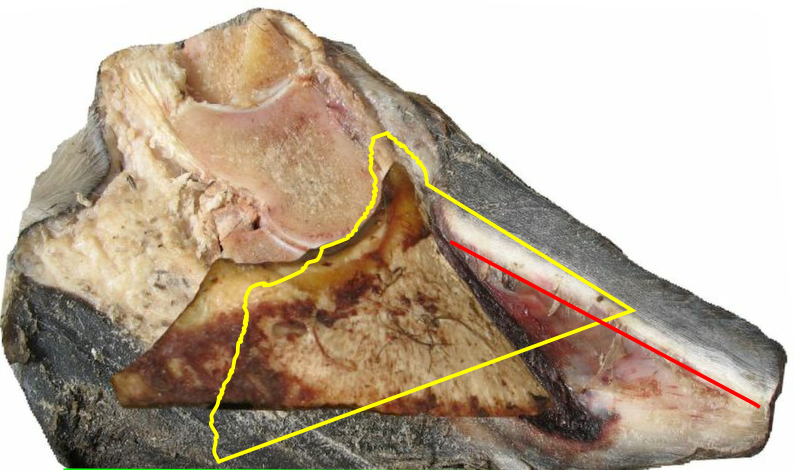
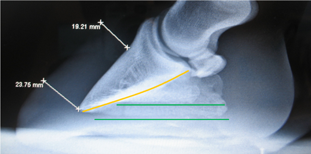
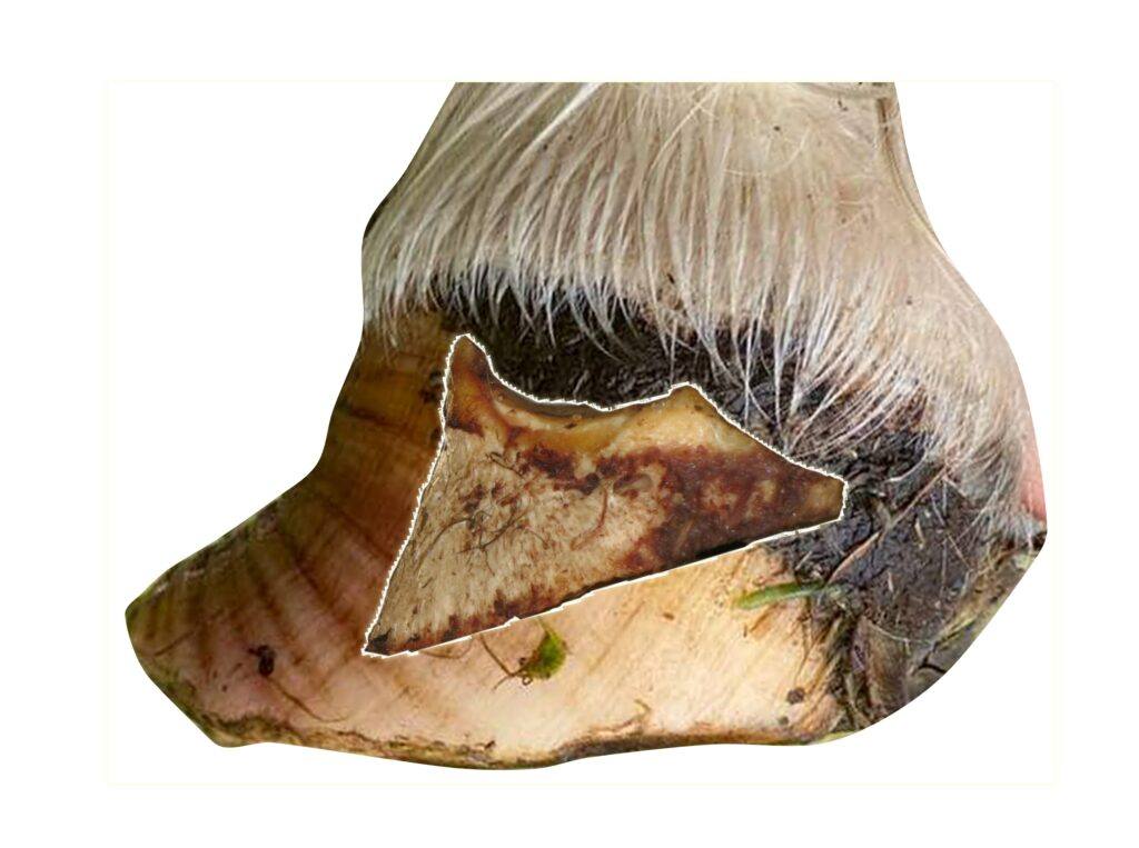
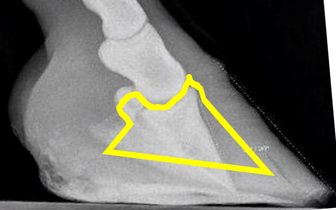
If the coffin bone was originally parallel to the toe wall, as the white marking suggests, and then “rotated” down to what the radiograph shows, where did all the materia in the red triangle go?

There is never any void inside the hoof where this missing tissue, or the coffin bone, could go. But the missing tissue must have gone somewhere, or?
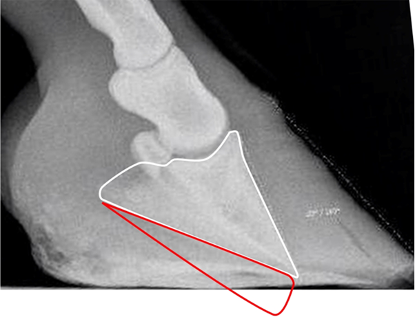
The answer is that it didn’t go anywhere because the bone has not moved. It was never up by the hoof wall; the hoof wall was down by the coffin bone, and got pressed up by ground pressure after suffering laminitis.
Why No Coffin Bone Has Ever Rotated
This is the point where interpretation ends and physical constraints take over.
The condition “Coffin Bone Rotation” is based on a 2-dimensional picture of a shadow. But hooves exist in four dimensions (they change over time), and biology requires space. When something truly moves, it must move somewhere, and that must be a flowing movement. It can’t disappear from one place and appear in a different place. The space it moved to must have been vacant, or the tissue that earlier occupied it must have gone somewhere, and that available space doesn’t exist.
That requirement alone is decisive.
If a rotation were to occur, the coffin bone and the navicular bone would rotate about the ball joint of the short pastern bone. The tip of the coffin bone would rotate in the direction of the yellow arrow and crush the soft, blood-filled sole corium while deforming the sole. Since this would mean working with an inverted leverage, it would demand a force equal to multiple times what the DDFT is capable of delivering, since the DDFT can only tip the hoof when the horse is moving forward, not making the hoof stand on its toe.

The pain from crushing the soft, blood-filled corium under the front part of the coffin bone would directly stop the horse from continuing. A crushed corium can’t be detected in a radiograph. The corium is clearly visible in a dissection, but a crushed corium has never been seen.

Rotating a coffin bone inside a hoof capsule must be considered an outrageous thought. It would have to destroy everything in there. The force needed would have to be immense, and if it had happened, there would be no chance for rehabilitation. The fact that the condition Coffin Bone Rotation is not just possible, but even easy to rehabilitate, is evidence that the traditional explanation for it is completely wrong.
Both the corium and the sole are usually intact and unharmed even after an alleged rotation. There is no crush damage under the coffin bone, but a lot of scar tissue between the bone and the hoof wall. This is evidence that the coffin bone has not rotated down, but something has happened above the coffin bone without the coffin bone moving.
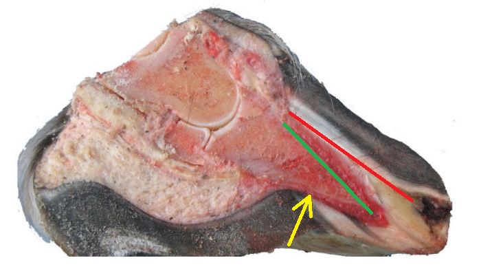
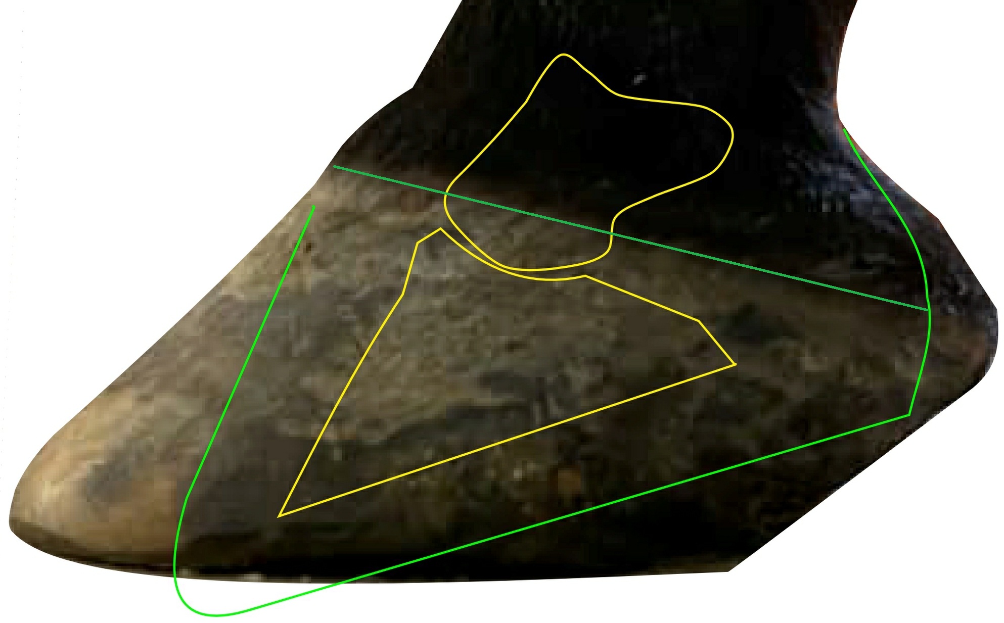
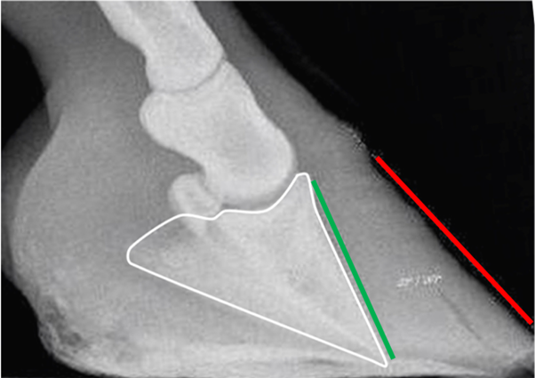
The red line indicates the alleged original angle of the coffin bone.
The green line indicates the current angle of the coffin bone.
The yellow arrow points to pristine sole corium.
The condition “Coffin bone rotation” does not imply that the coffin bone has rotated.
It only means that the toe wall and the coffin bone are no longer parallel. The “rotation” is an alleged explanation, not a part of the condition.
All deformed laminitic hooves have the condition Coffin Bone Rotation because that is exactly what it means: the hoof wall is no longer parallel to the coffin bone. Some of them also got a stretched sole when the laminitic hoof wall was pushed forward by ground pressure. If the laminar junction is in good condition, the hoof wall will pull and stretch the sole when pushed forward, but if it has been weakened, it will break instead. A sole that has become stretched past the hoof wall corium is evidence that the hoof wall changing angle, not the coffin bone rotating, was the reason for the creation of the condition Coffin Bone Rotation.
All laminitic hooves that get a steeper “healing angle” at the top of the hoof wall during rehabilitation are evidence that it was the laminitic hoof wall that caused the condition Coffin Bone Rotation, not a rotating coffin bone. The healing angle is created when the toe has been shortened enough to reduce ground pressure on the toe wall, which lets the new hoof wall naturally follow the coffin bone when growing down. The old and deformed toe wall, with its scar horn, will still be present until gradually trimmed away. If the coffin bone had been re-rotated, there would not have been such a reset of the toe wall angle.
Some radiographs are not evidence for or against the traditional rotation theory, but they can explain why the theory came to be. But basing a theory only on an observation that merely makes it possible is not scientific.
Traditional rehabilitation of Coffin Bone Rotation means pressing the coffin bone back up to the hoof wall. This is, however, impossible since that space is now occupied by scar horn filling all the volume needed for the coffin bone to be re-rotated into.
The Sole Corium Reveals What Has Happened
A radiograph is a shadow.
A photograph is 2-dimensional.
Physics happens in 4 dimensions (length, width, height, time)
When we look at where the rotating coffin bone is said to have passed, from its original position parallel to the hoof wall down to the sole, on a radiograph, we see nothing. On a cross-section, we see pristine tissues where there should have been total disorder after the passing coffin bone.
In hooves diagnosed with “coffin bone rotation,” the corium and sole beneath the coffin bone remain present and continuous. Nothing has disappeared. Nothing has been compressed out of existence or even compromised.
This alone is evidence that the bone has not rotated downward.

Blue marking = alleged natural position
Yellow marking = current position
Black marking = volume the coffin bone alleged has been passing through
Green marking = Where the remains from the black marking should be today
There is no crush damaged tissue visible anywhere regardless of the above.
What has changed is the relationship between the hoof capsule and the bone, not the bone.
Radiographs show angles, not mechanisms.
The diagnosis is always based on one of two observations:
- The distance measured from the outside of the toe wall to the coffin bone is not the same, high and low.
- The angle of the outside of the hoof wall is not the same as the toe angle of the coffin bone.
That observation is real, and if the conditions are fulfilled, something is wrong.
But it does not prove bone movement.
It shows only that the relation between the hoof wall and the coffin bone has changed.
When laminitis weakens lamellar attachment, the capsule becomes loose and can migrate, flare, and deform under ground pressure. The bone, constrained by anatomy and supported from below, does not.
This is why the condition can disappear when hoof capsule geometry is restored — without anything changing to the bone itself.
Rotating a bone back is physically impossible; instead, you remove the forces that deform the capsule and allow the hoof to rebuild around the existing skeletal position.
Rehabilitation occurs by natural regrowth, not by human intervention. That is why real rehabilitation is possible.
The key mechanical conclusion
If “rotation” disappears when the hoof capsule geometry is restored (toe leverage and wall position), then the visible “rotation” was produced by the capsule’s deformation—not by a bone pivot.
How much a laminitic hoof deforms from ground pressure depends entirely on hoof shape.
A hoof with long toes will suffer severe deformation, while a perfectly shaped hoof with short toes might not suffer any permanent deformation at all. Yes, that is correct: founder, laminitis, no deformation, and the pony is running again in four days. The reason this is not common knowledge is that perfect hoof shape is very rare.
A laminitic trim is not nearly as mystical or complicated as some claim. It is only about removing unnatural leverage by shortening the toes and lowering the heels to relieve ground pressure on the toe wall.
Exactly how to determine how much can and should be removed will be explained later in a dedicated chapter on laminitis trimming. This will also include completely safe but extreme toe shortening.

Once the hooves of your laminitic riding horse/pony have been trimmed to this form, you should already be riding again. There is no risk for the hooves in beginning to ride at this stage — everything that could cause harm has already been removed. There is no need for a long recovery rest. I know some people say that recovery takes 9 months, and that might be right if you don’t trim the hooves the right way. Hooves like this must be trimmed after their own schedule, 1-6 weeks, to prevent ground pressure on the hoof wall from increasing again. This means that nailed-on shoes and glue-on boots are out of the question.
Summary of evidence
- Ground parallel coffin bones with Coffin Bone Rotation should be impossible since the coffin bone can’t be rotated and ground parallel at the same time.
- Since the space between the hoof wall and the coffin bone directly fills with wound serum that hardens to scar horn, rehabilitation must be impossible, which it’s not.
- When you replace the half coffin bone you can see in radiographs, or cadaver hooves cut lengthwise, with a complete coffin bone, you will see that if you place it parallel to the toe wall on a hoof that suffers the condition coffin bone rotation, the bone will not fit in the hoof capsule, have an extreme negative angle, or it might be ground parallel. All three defy the rotated coffin bone theory.
- The connection point of the DDFT and its angle demands an unrealistic force to be able to rotate the coffin bone.
- There is no space anywhere for the coffin bone to rotate into.
- The sole corium under the coffin bone is undamaged in the hooves with coffin bone rotation.
- It is physically impossible to rotate the coffin bone inside the hoof capsule while the horse is standing on it.
- That the new hoof wall grows parallel to the coffin bone during rehabiliation prooves that it was the hoof wall that migrated during the “rotation”.
Conclusion
- Traditional hoof care is based on the misconception that the coffin bone is suspended from the laminae of the hoof wall. This would break the laws of nature.
- The theory of the rotating coffin bone must be considered completely unscientific, as it violates basic physics and the laws of nature.
- The condition coffin bone rotation says nothing about the coffin bone’s orientation compared to the horizontal plane, only the parallelity to the toe wall.
- Dissections prove that the coffin bone can’t have rotated down since the sole corium is not damaged.
- If the coffin bone didn’t rotate down, it must have been the hoof wall that rotated up to create the void between them.
- It is the heel height that affects the angle between the coffin bone and the ground. Nothing else.
- When you understand that the problem is capsule mechanics, not a rotating bone, “coffin bone rotation” stops being a fatal label and becomes a solvable mechanical situation.
- “Coffin Bone Rotation” is easy to rehabilitate, and there is no reason to fail.